
Correlating STED and synchrotron XRF nano-imaging to explore metal functions in synapses
AIP Conference Proceedings have published an article by Domart et Al which in which researchers “correlate stimulated-emission-depletion microscopy (STED) of proteins and synchrotron X-ray
fluorescence (XRF) imaging of trace metals, both performed with 40 nm spatial resolution, on primary rat hippocampal neurons.”
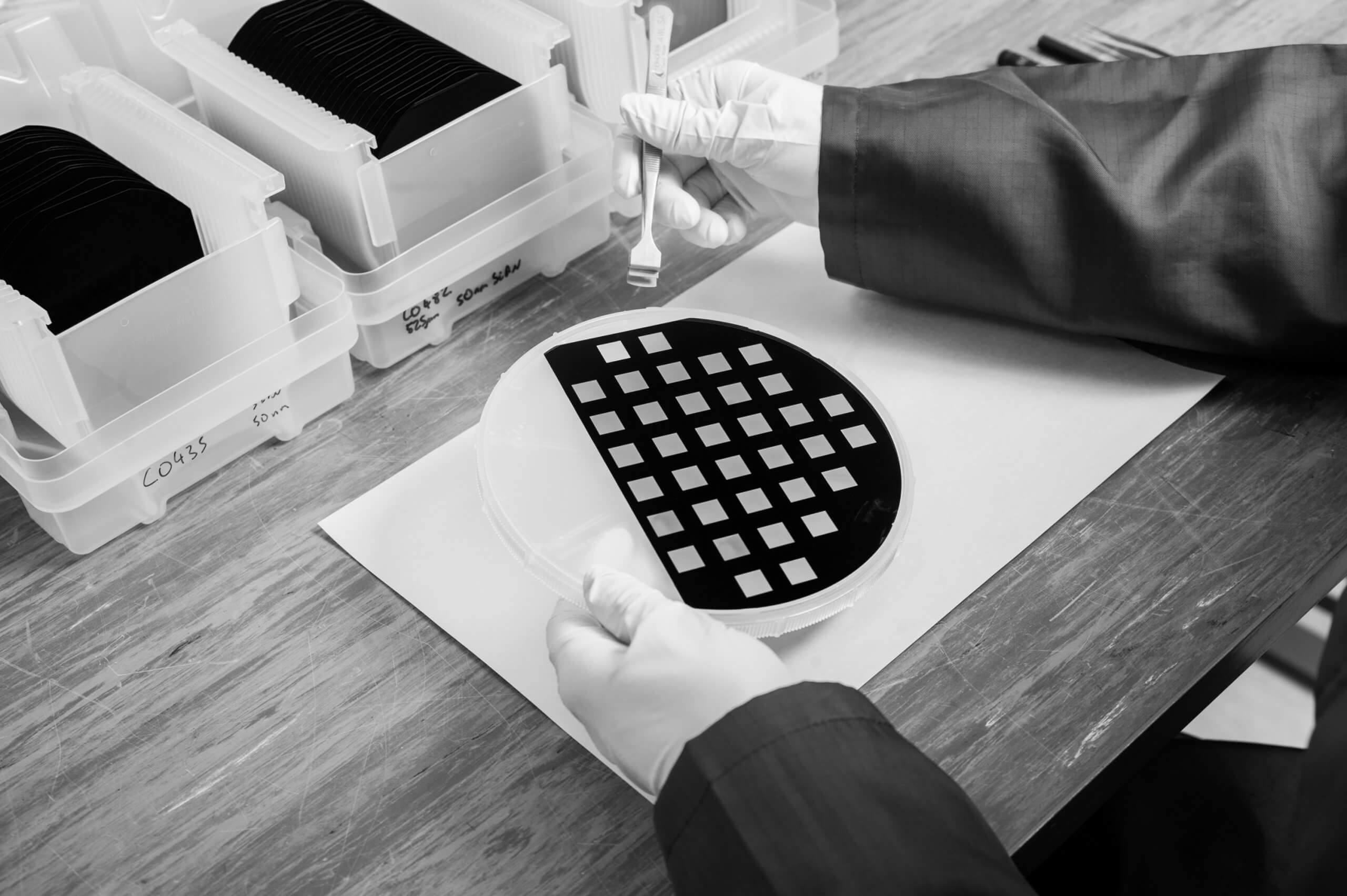
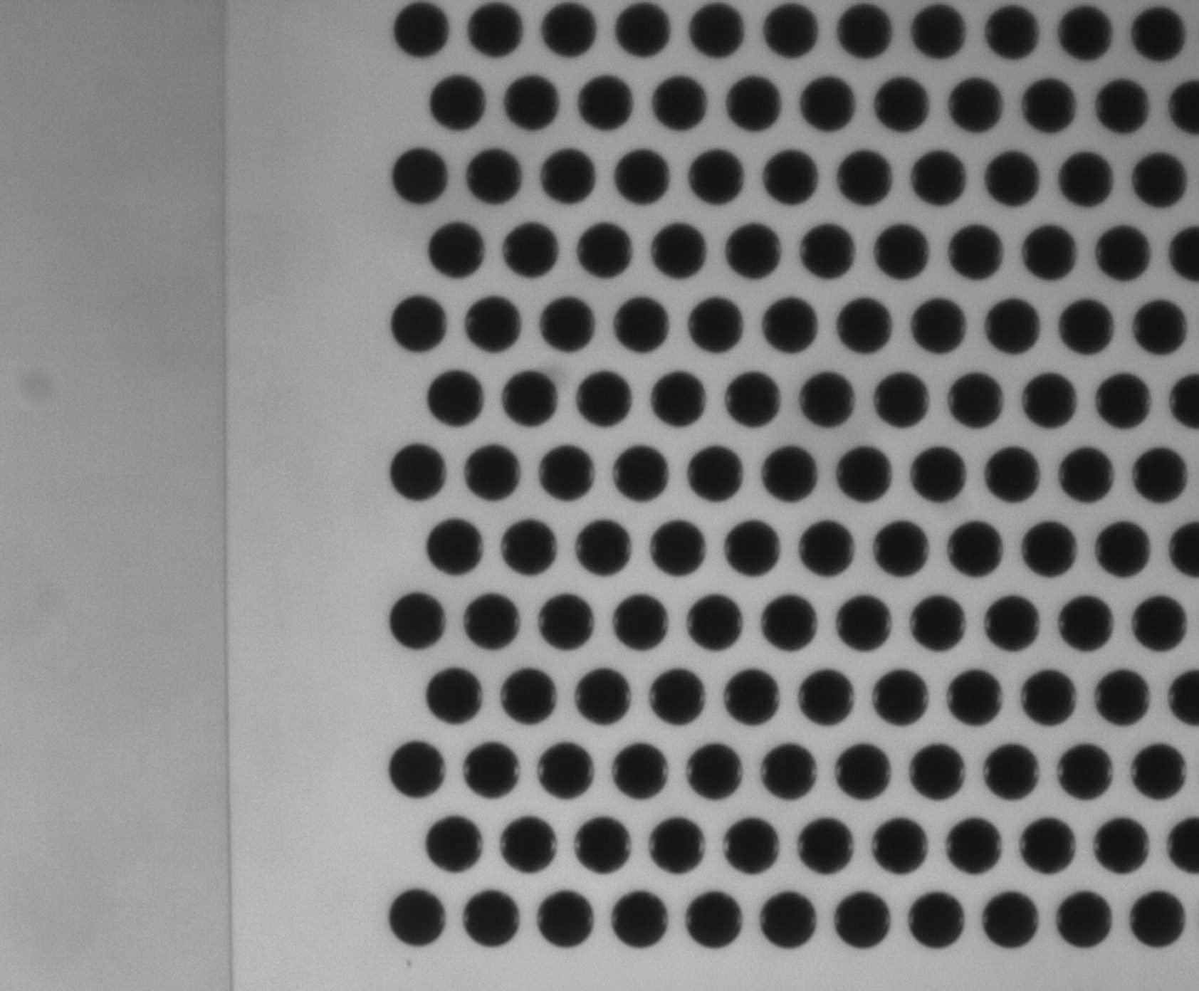
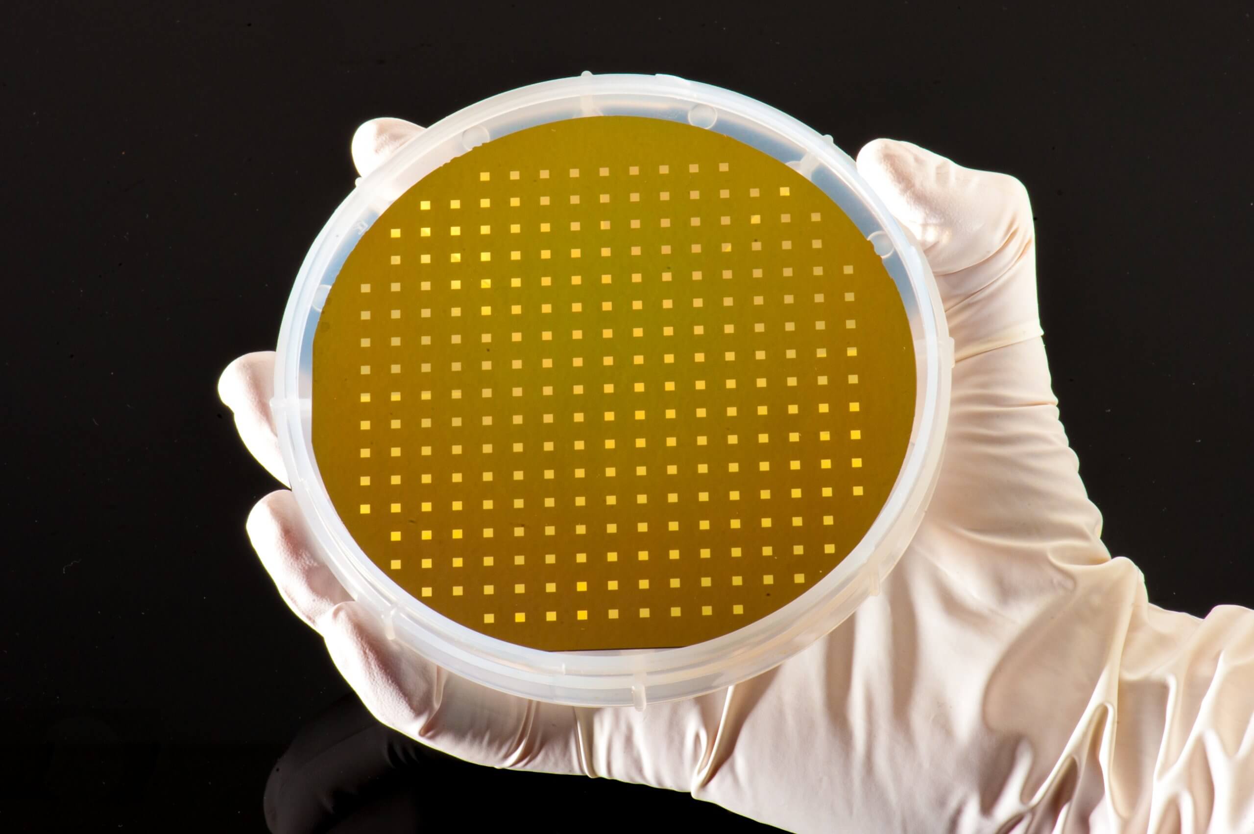
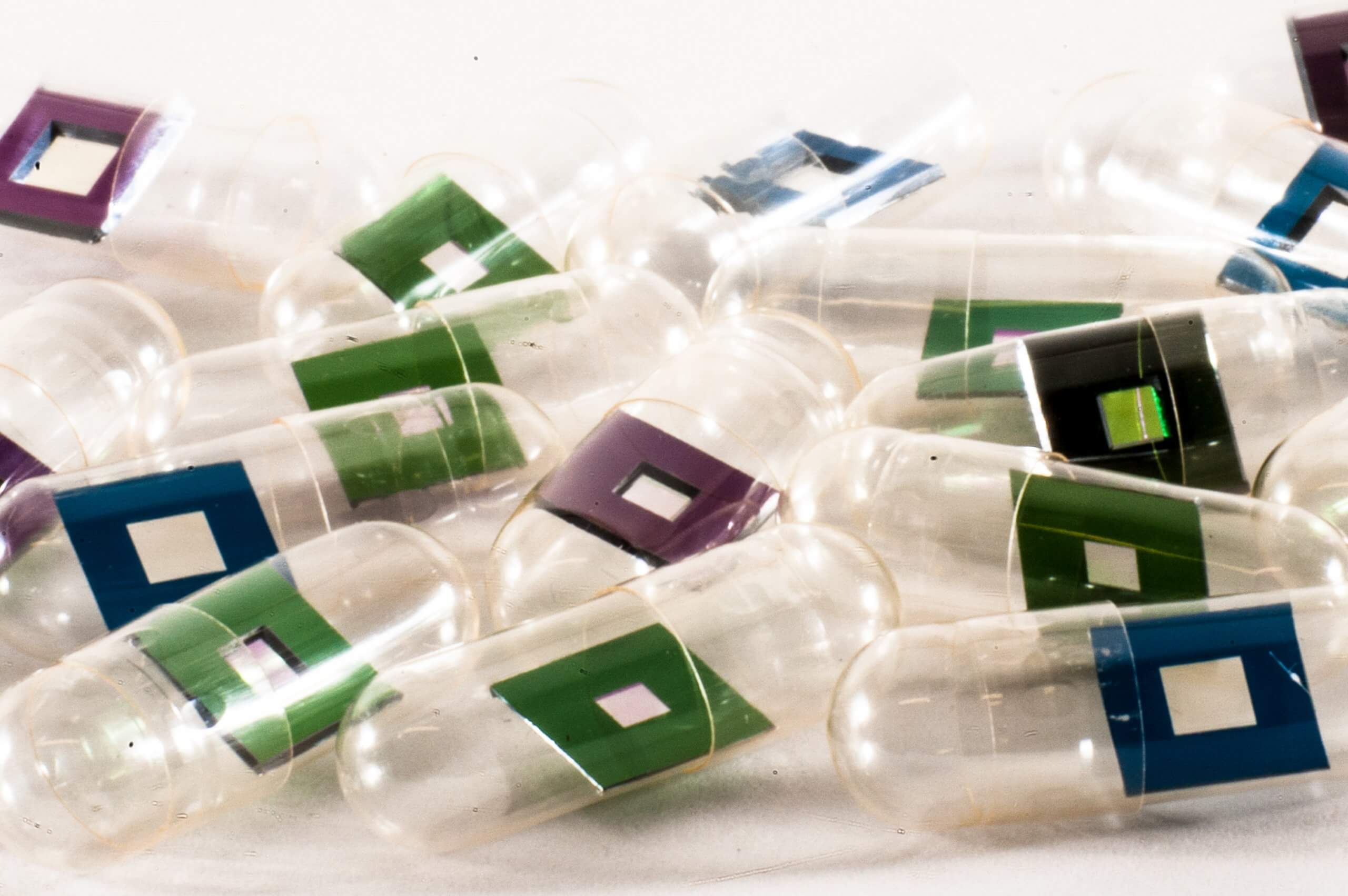
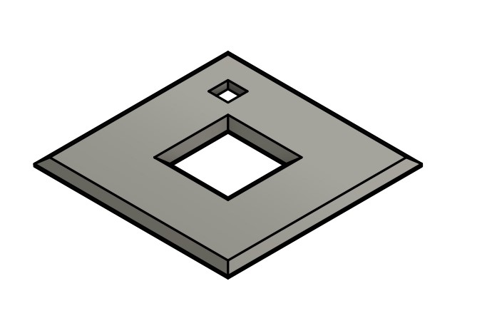
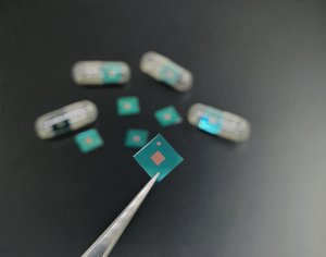
Primary rat hippocampal neurons were cultured on Silson’s silicon nitride orientation membranes of frame size 5 x 5 mm² and thickness 200 µm and membrane size 1.5 x 1.5 mm² and thickness 500 nm. During our manufacturing process, we add a smaller membrane in one corner of the chip for orientation purposes:
Nano-SXRF imaging was then performed on the ID16A Nano-Imaging beamline at the European Synchrotron Radiation Facility (Grenoble, France). Their conclusion ends with: “From a methodological perspective, the combination of STED super resolution microscopy and nano-SXRF imaging opens numerous perspectives of application to investigate the chemical biology of metals.”
Follow this link to read the entire article!