Case studies

ChemRxiv has published an article by Ortega et Al about the study of cellular functions of essential metals. The abstract reads “we correlate protein localization, using fluorescence light microscopy (FLM), and metal distribution with synchrotron X-ray fluorescence (SXRF), a high-sensitivity and high-spatial-resolution technique for metal imaging”
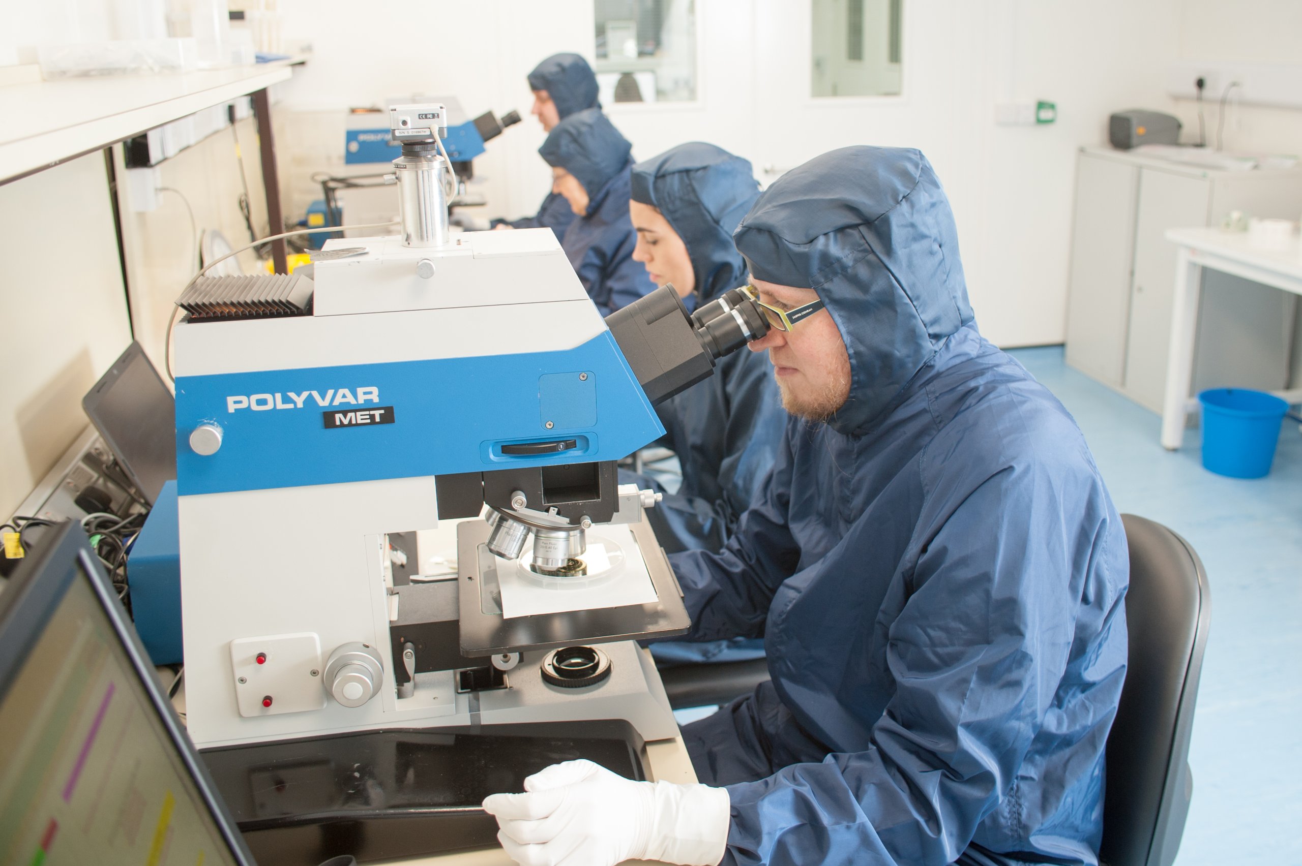
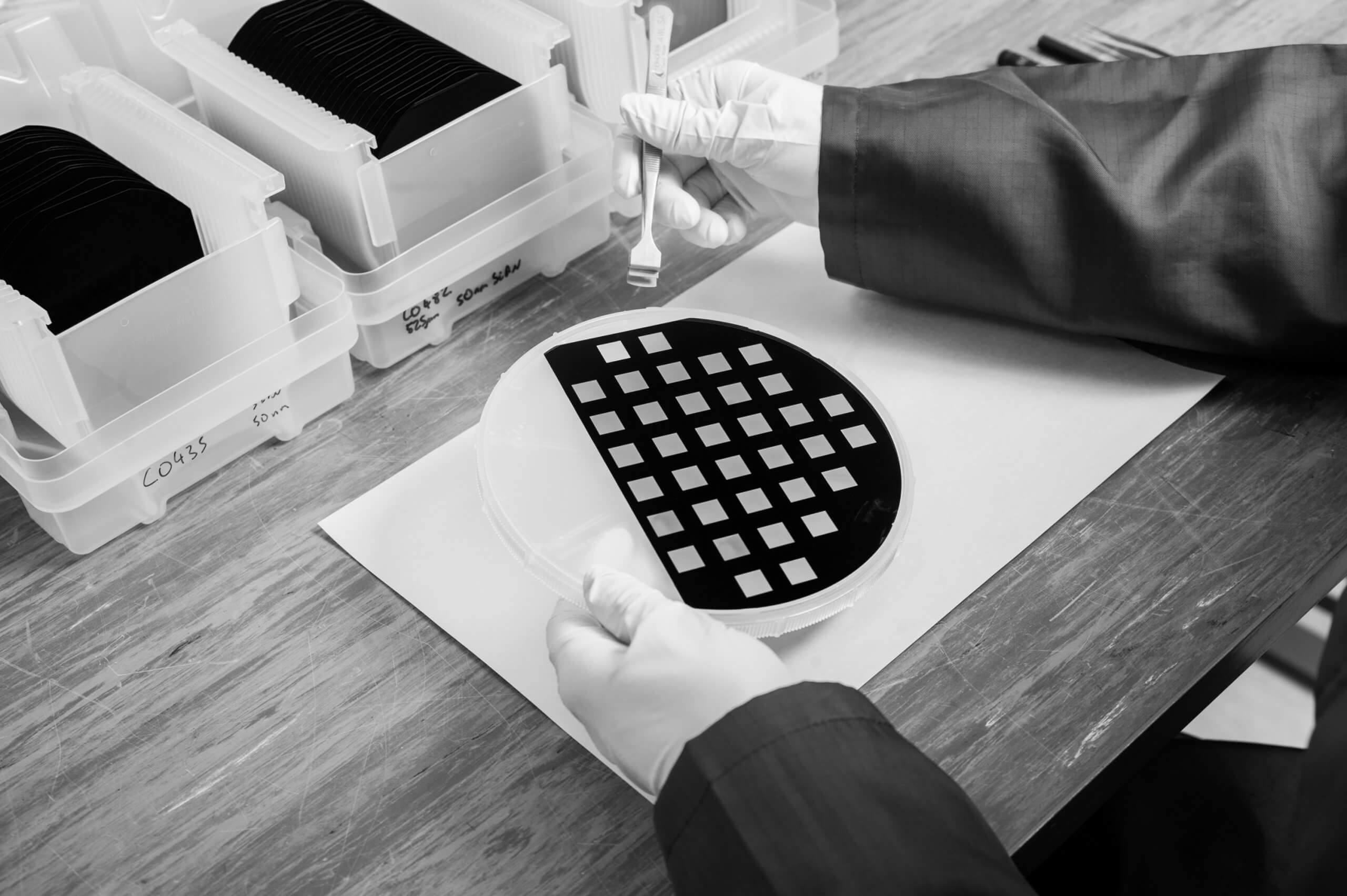
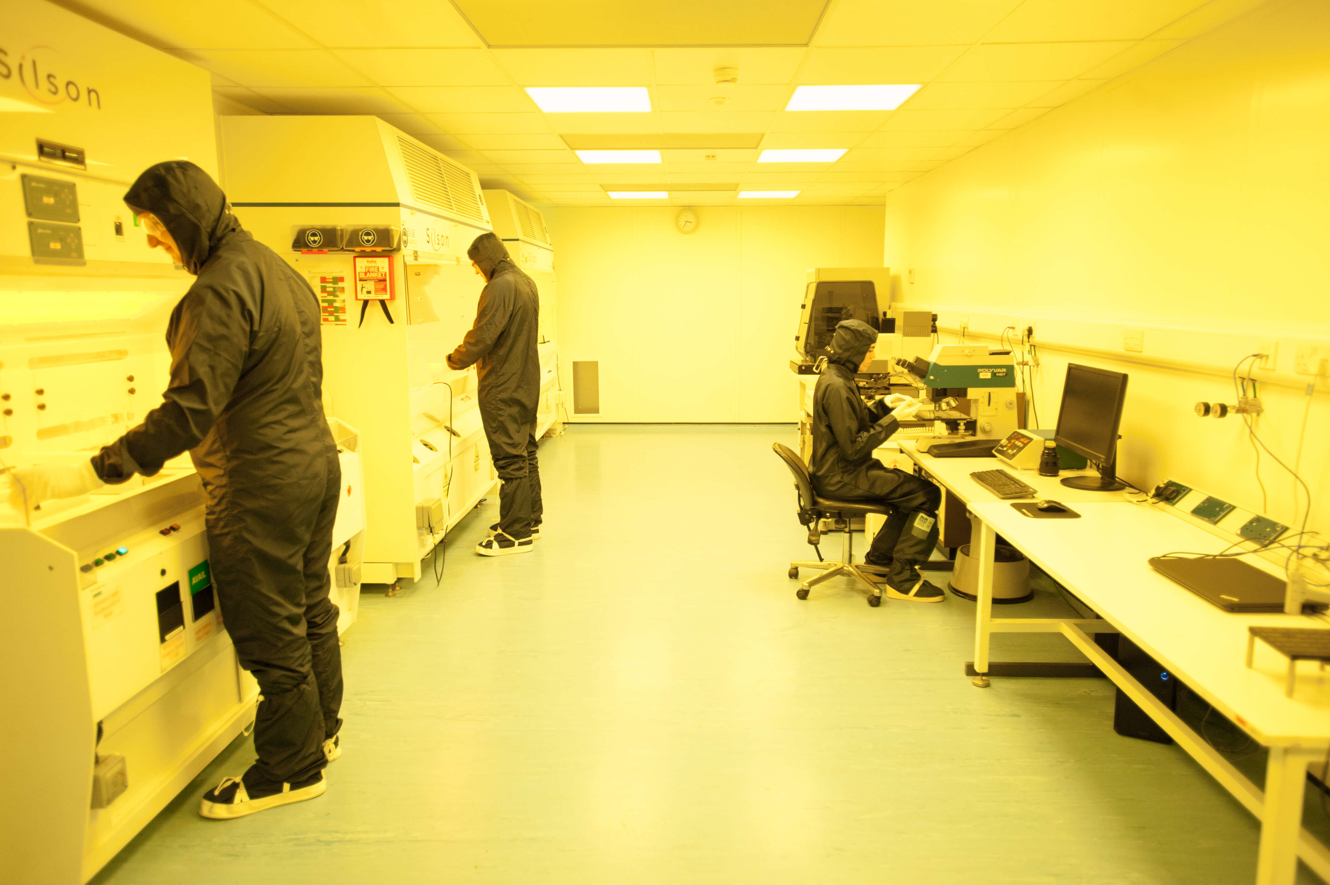
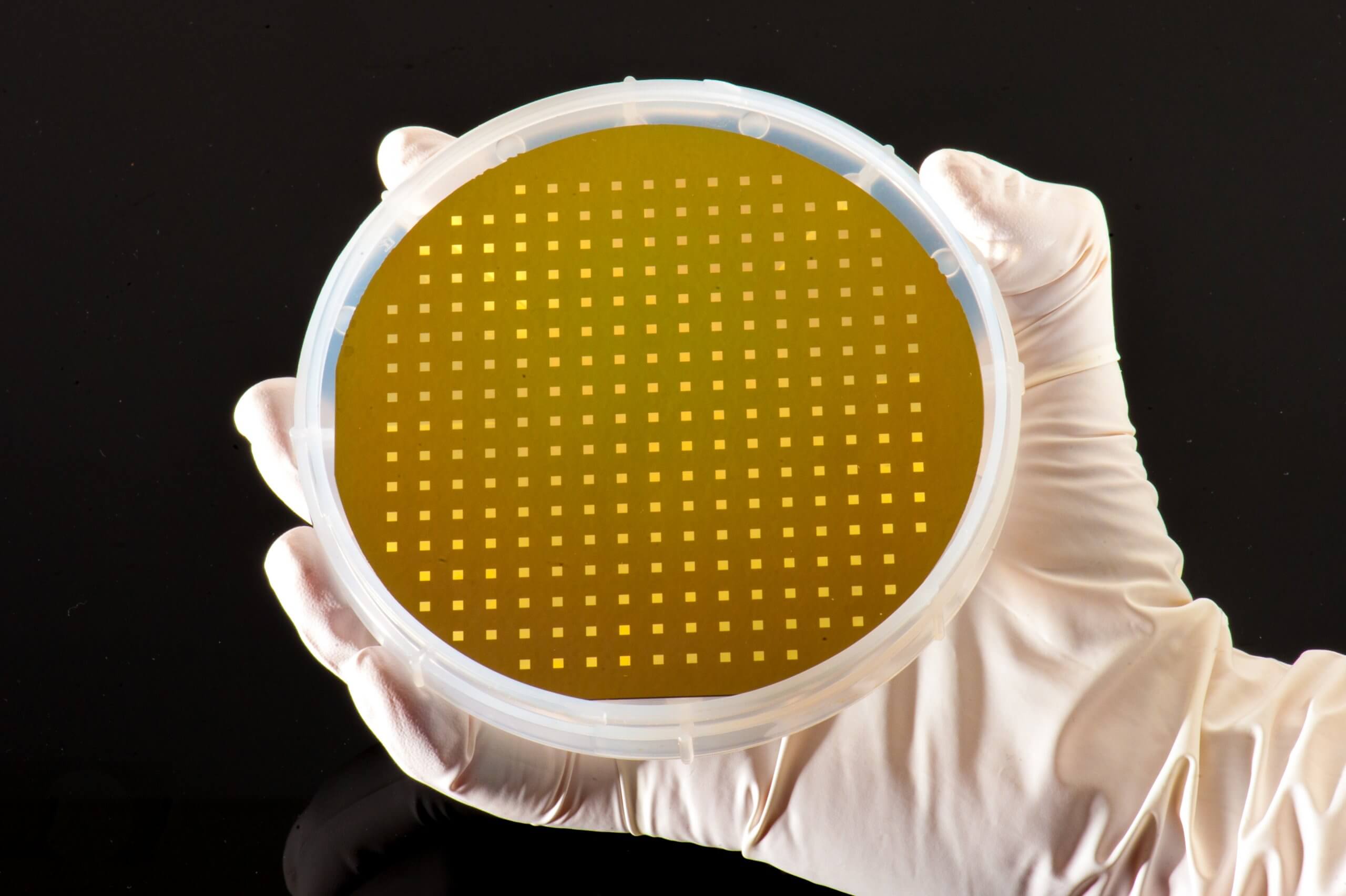
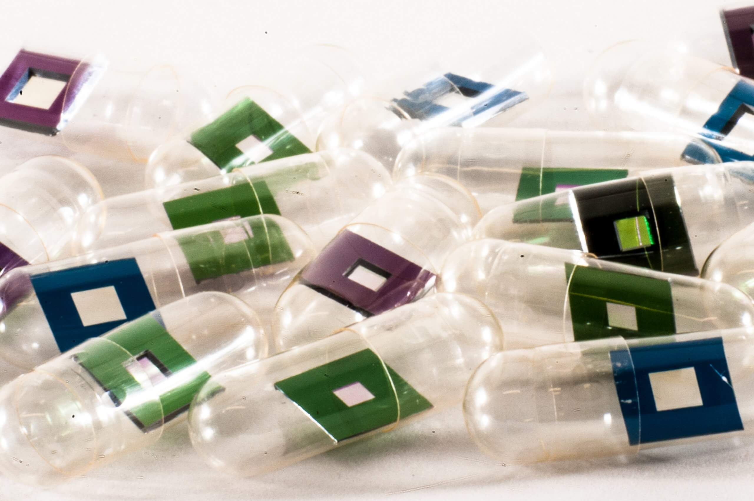
Both of the imaging techniques mentioned above were performed under cryogenic conditions. Silson’s silicon nitride orientation membranes of frame size 5.0 x 5.0 mm and thickness 200 µm and membrane size 1.5 x 1.5 mm and thickness 500 nm were used as the cell culture substrates for the SXRF analyses. Our membranes used here also have a smaller membrane in one corner of the frame, for orientation purposes.
Follow this link to read the full paper!