Case studies

Article published by Ortega et Al at the University of Bordeaux in which they have developed a correlative nano-imaging approach to address the challenge of understanding the role of metals on synaptic functions.
Their approach appears successful as it “allows for ultra-sensitive detection of trace metals using highly focused synchrotron radiation beams” and the abstract concludes that they have provided “proof-of-principle for correlative imaging of metals and proteins at the synaptic scale and discuss[ed] the present limitations and future developments in this area”.
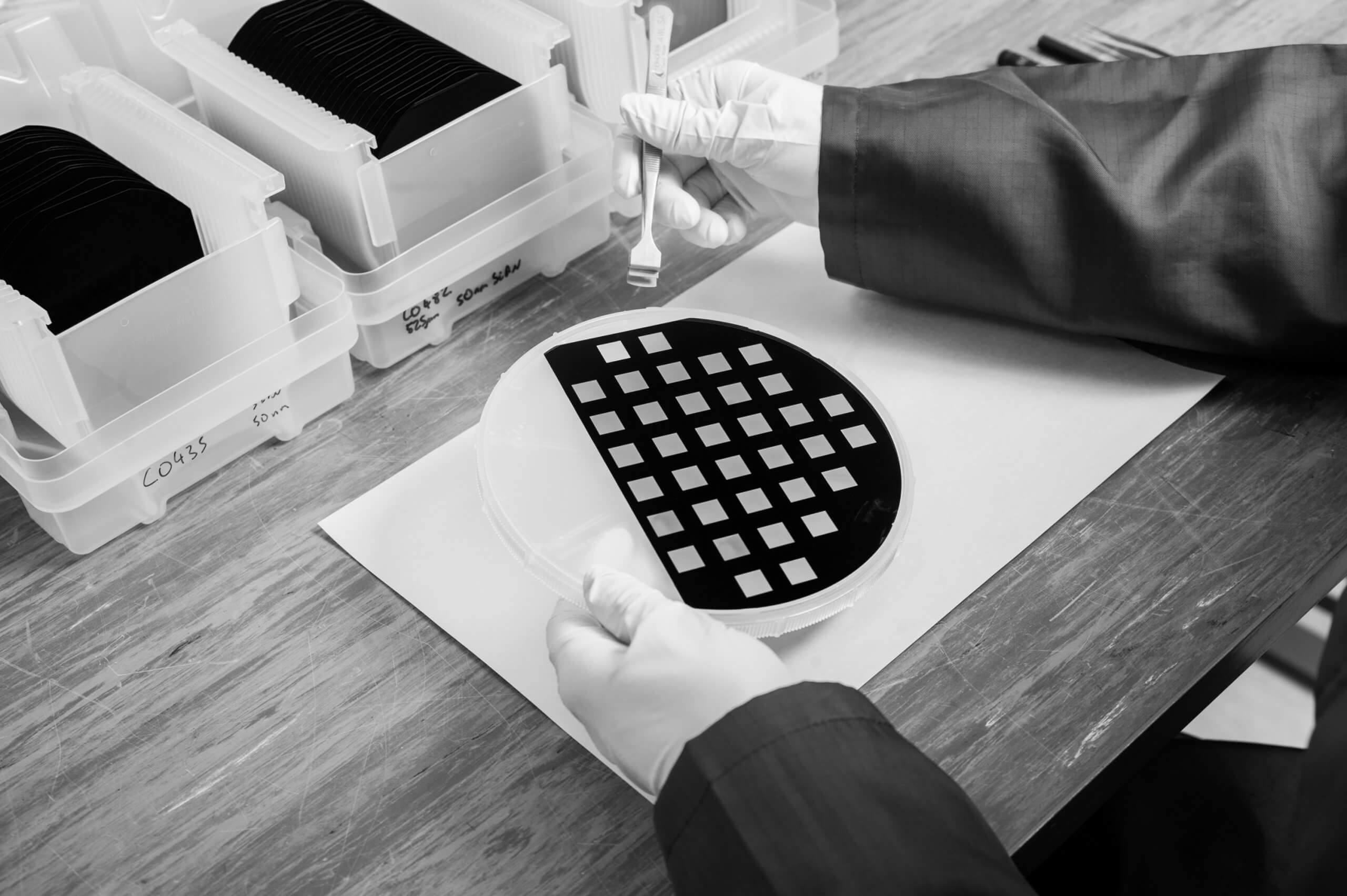
Silson’s silicon nitride orientation membranes were used in the synchrotron X-ray spectro-microscopy experiments (frame size of 5 mm× 5 mm, and a frame thickness of 200 µm, membrane size of 1.5 mm × 1.5 mm, and a membrane thickness of 500 nm). The membranes were manufactured with a marking on the outer edge, which is used for orientation purposes during experiments.
Click here to read the full article!