Case studies

Research published by Pucetaite et Al, investigates micro-to-nanoscale fungal-mineral interactions in soil fungi. The abstract reads: “In this work, we have grown saprotrophic and symbiotic fungi in contact with two soil minerals with contrasting properties: quartz and goethite, on top of X-ray transparent silicon nitride membrane windows and analyzed fungal hyphae by synchrotron-based scanning transmission X-ray microscopy in combination with near edge X-ray fine structure spectroscopy at C(K) and Fe(L) absorption edges.”
The research “demonstrates the link between processes initiated at the single-cell level to macroscale phenomena. Thus, spatially resolved chemical characterization of the microbial–mineral interfaces is crucial for an increased understanding of overall carbon cycling in soil.”
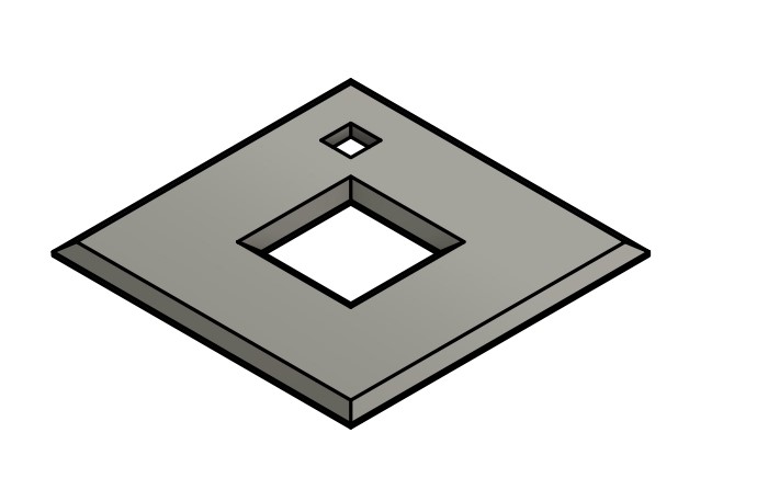
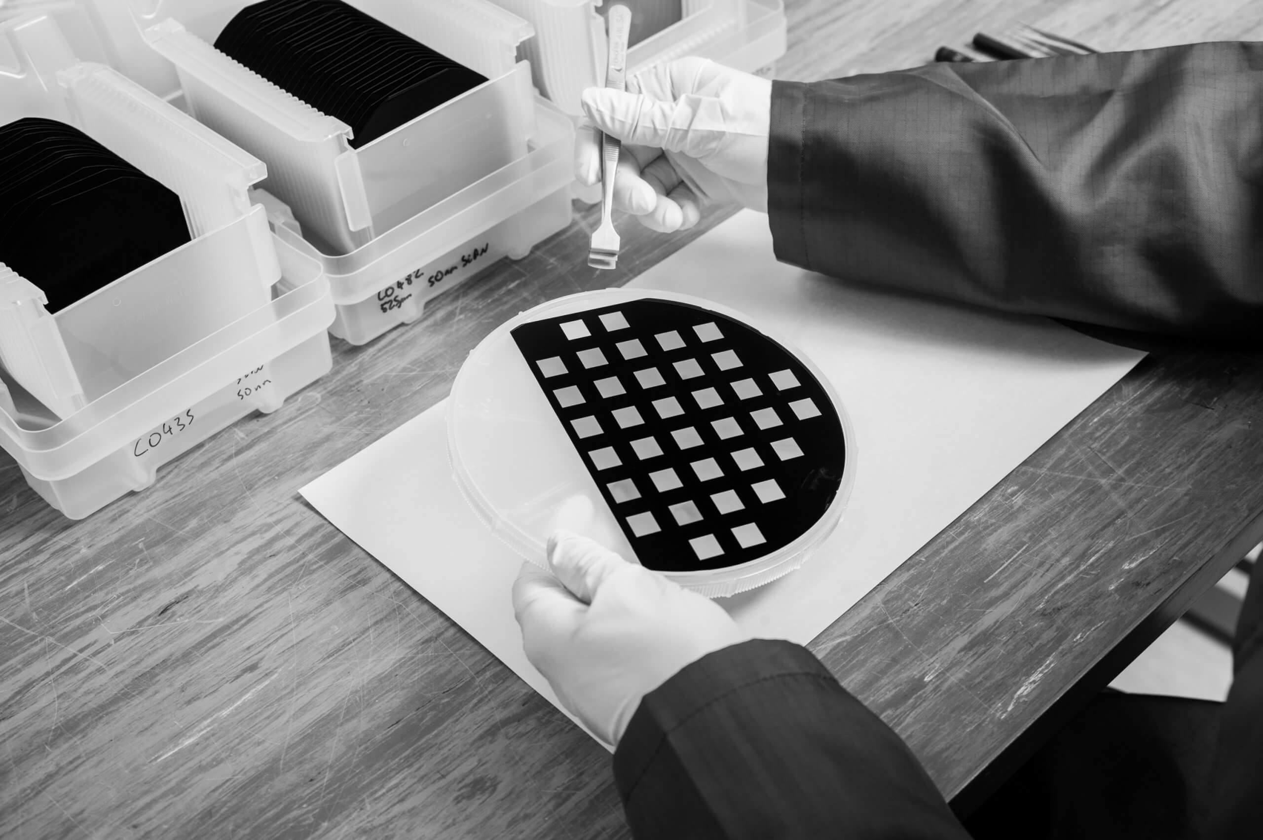
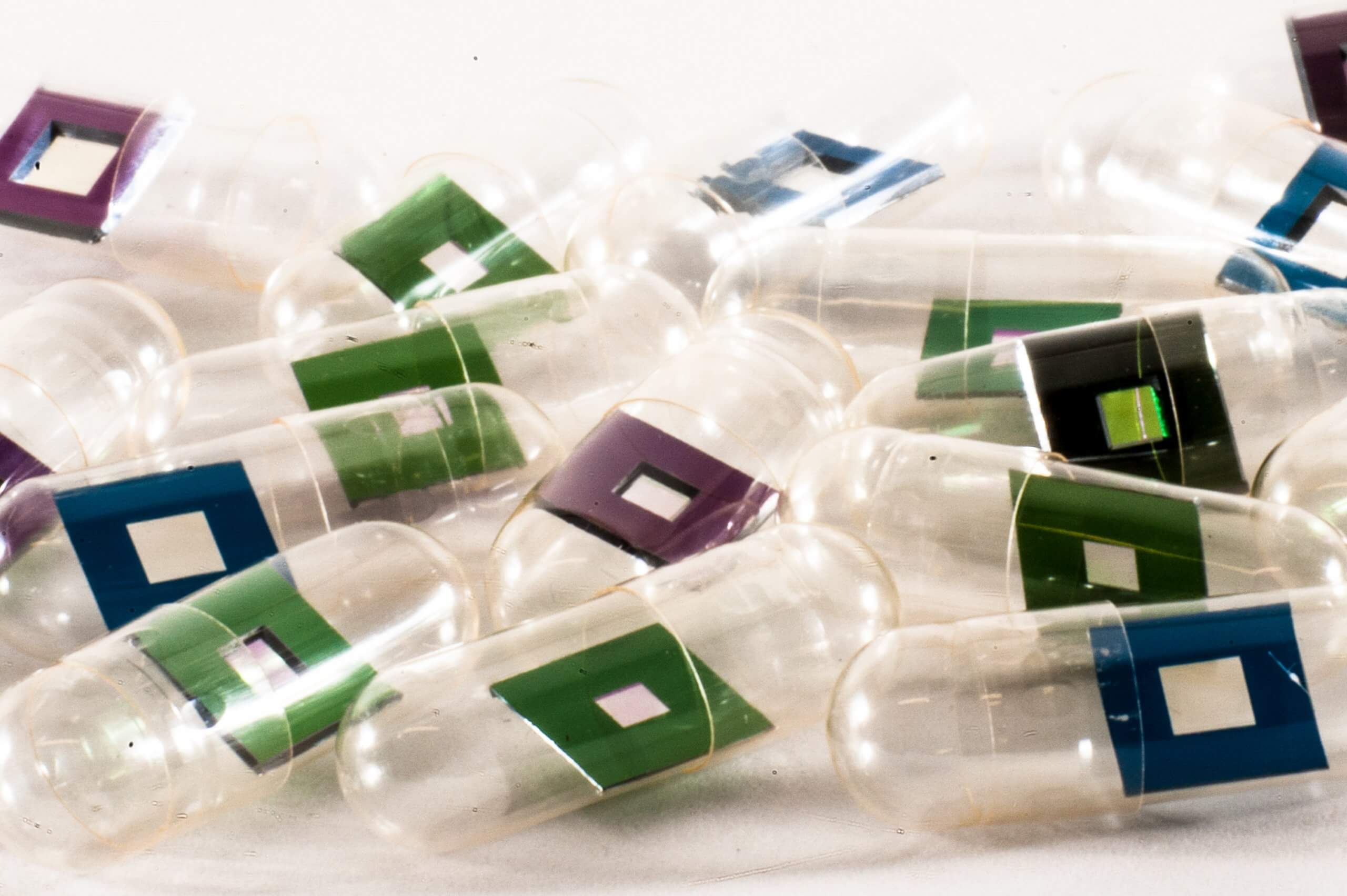
Silson’s membranes were used for the sample preparation. The sizes used were membrane dimensions of 75 nm in thickness and 500 × 500 μm or 750 × 750 μm in size, and frame dimensions of 200 μm in thickness and 5 × 5 mm or 2.75 × 2.75 mm in size, depending on the sample holder type used in the particular synchrotron facility.
Here is the link to read more about this research!