Case studies

The American Chemical Society has published an article by Byrnes et Al in which “micro- and nanoscopic X-ray techniques were used to investigate the relationship between uranium (U) tissue distributions and adverse effects to the digestive tract of aquatic model organism Daphnia magna [a freshwater invertebrate] following uranium nanoparticle (UNP) exposure.”
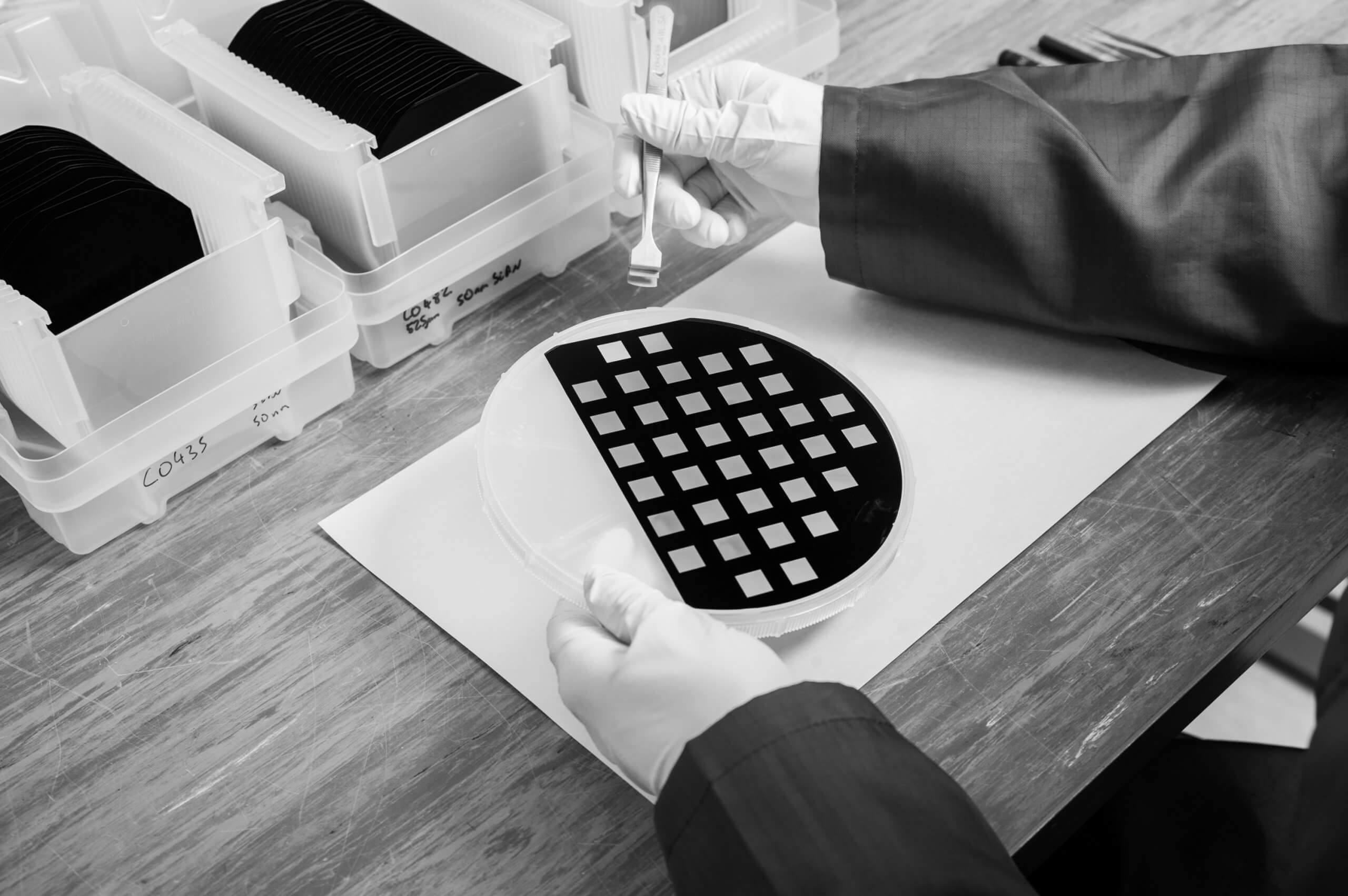
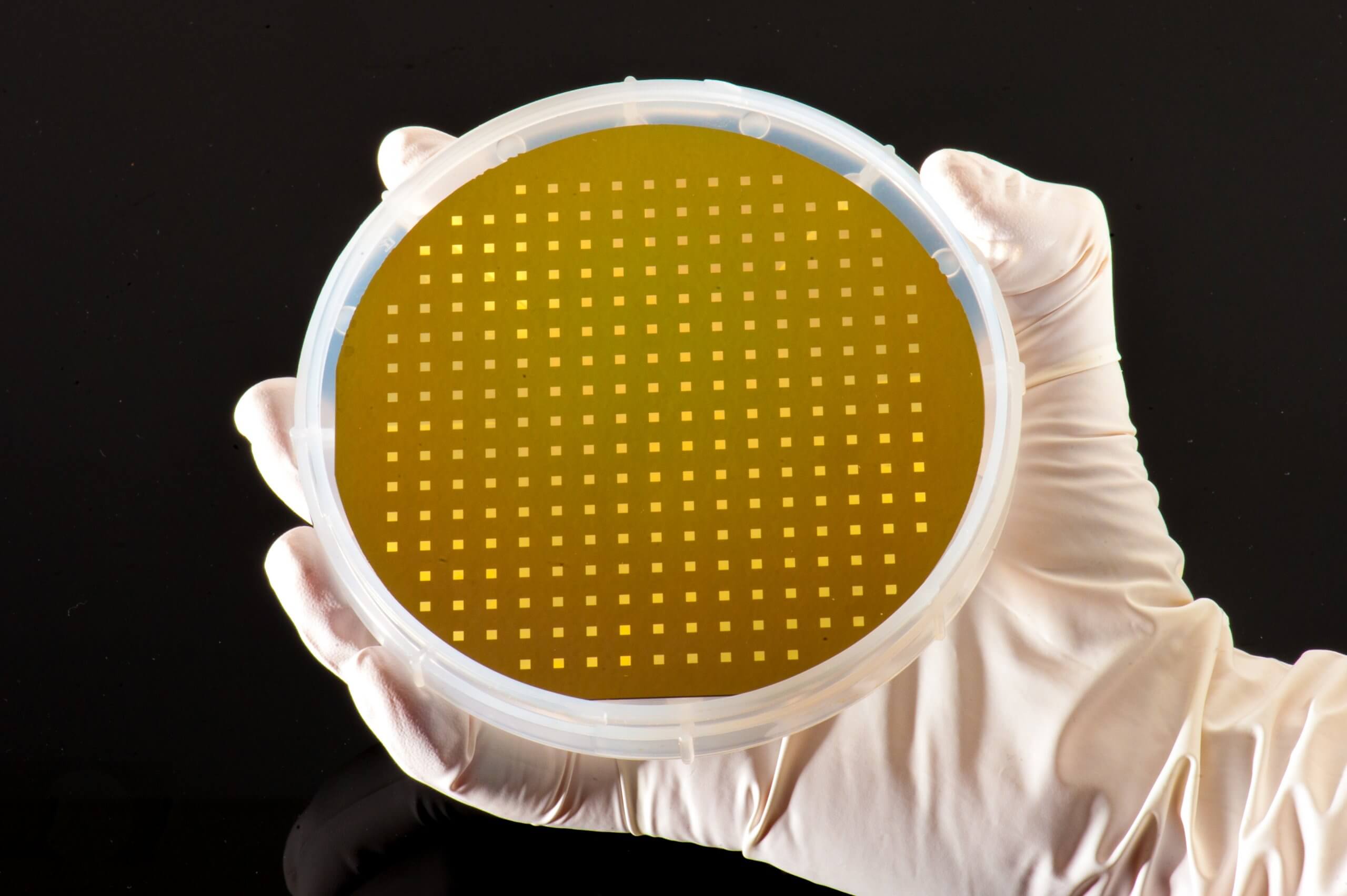
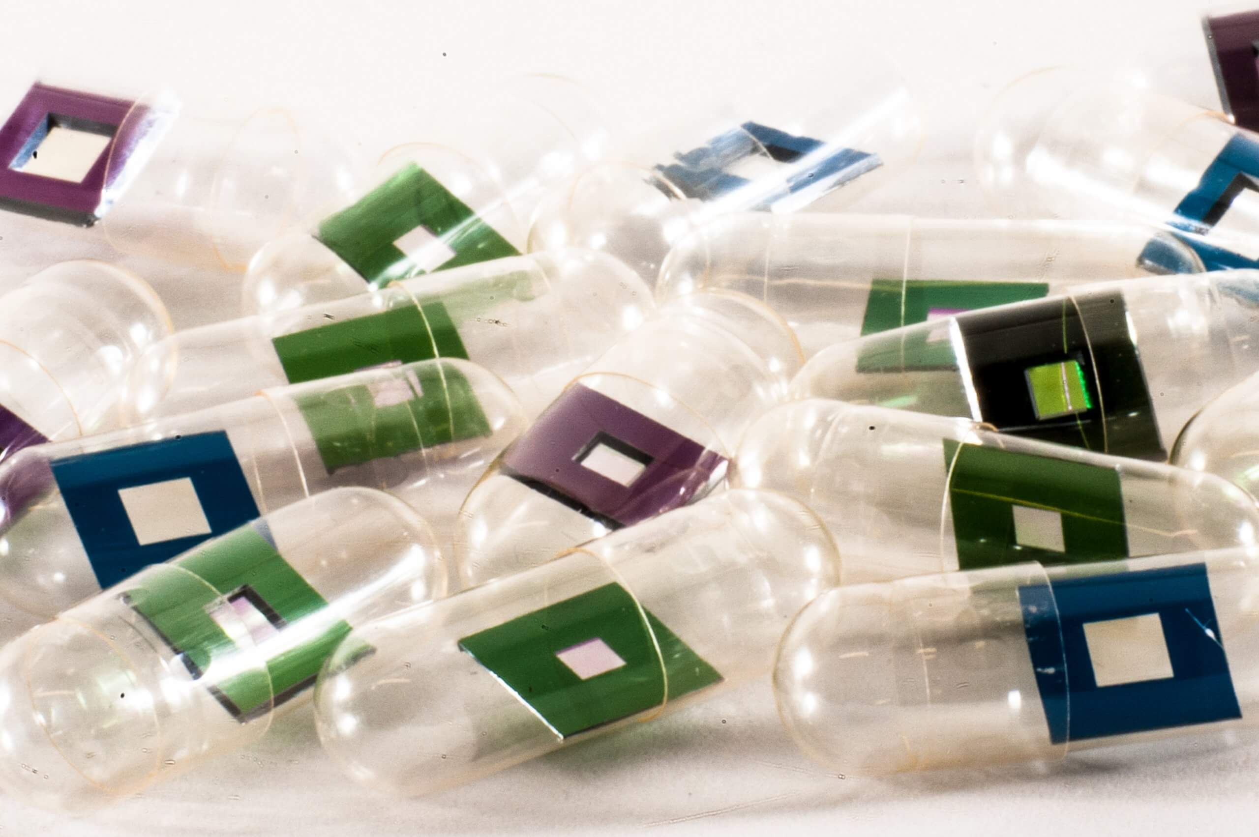
For the synchrotron-based nanoscale x-ray fluorescence imaging (nano-SRXRF), samples were mounted onto Silson’s 5×5 mm^2 silicon nitride membranes, and the x-ray fluorescence scanning was conducted at the I14 Hard X-ray Nanoprobe beamline of the Diamond Light Source. Also, further analysis of “daphnid midgut tissues were conducted using scanning transmission electron microscopy (STEM) with energy-dispersive X-ray spectroscopy (EDS)”.
Click here to read the article in full!