Case studies

Biomedical Optics Express have published an article by Wittmeier et Al, a study in which they “combine and correlate hard X-ray propagation-based phase contrast tomography and visible light confocal microscopy in three dimensions to probe DNA in whole cell nuclei of NIH-3T3 fibroblasts”, in order to visualize substructures within the cell in a complementary manner.
The research “enables the quantification of the electron density, volume and optical fluorescence intensity of nuclear material. By joining all of this information, we are able to spatially localize and physically characterize both active and inactive heterochromatin, euchromatin, pericentric heterochromatin foci and nucleoli”.
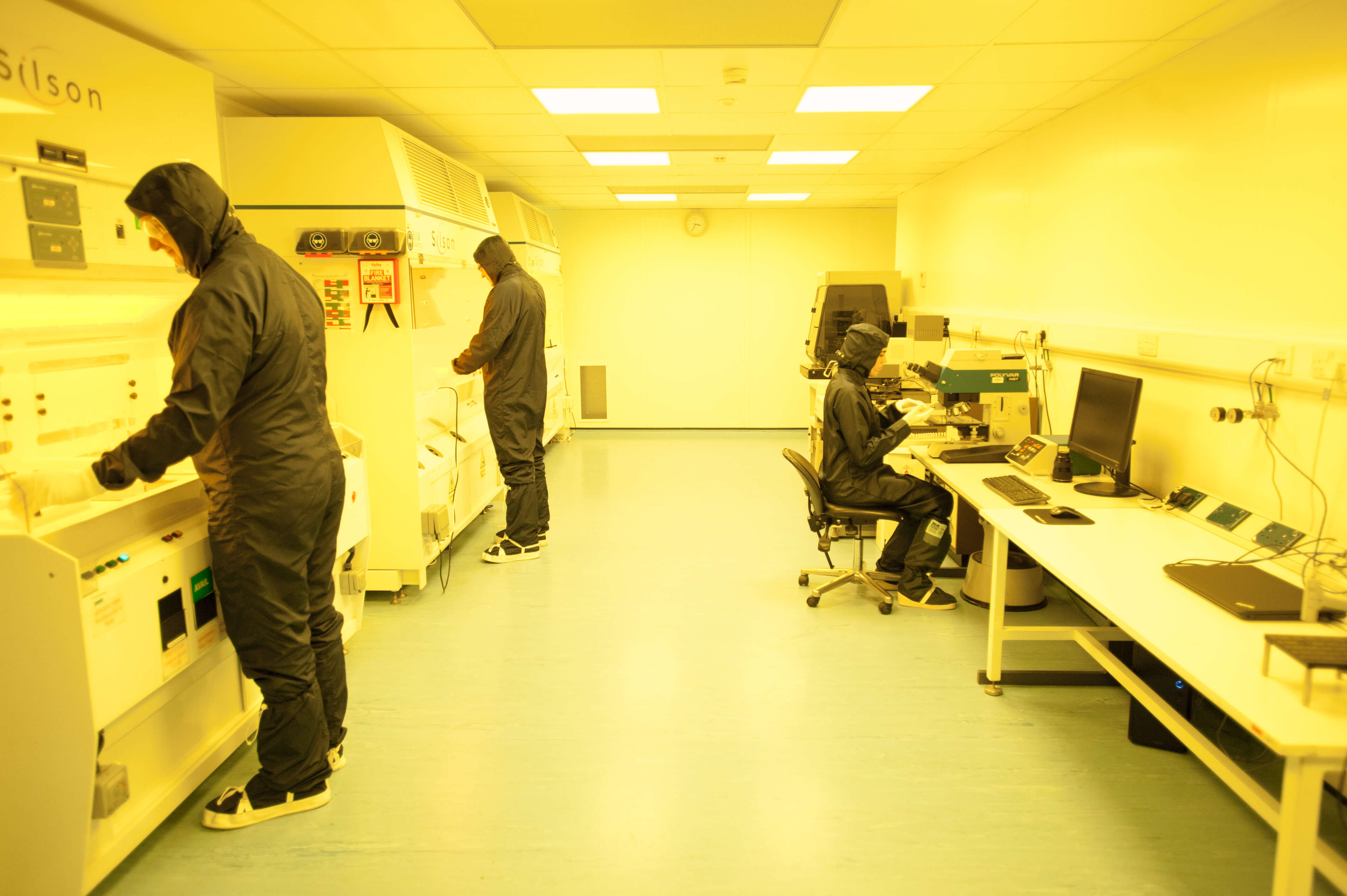
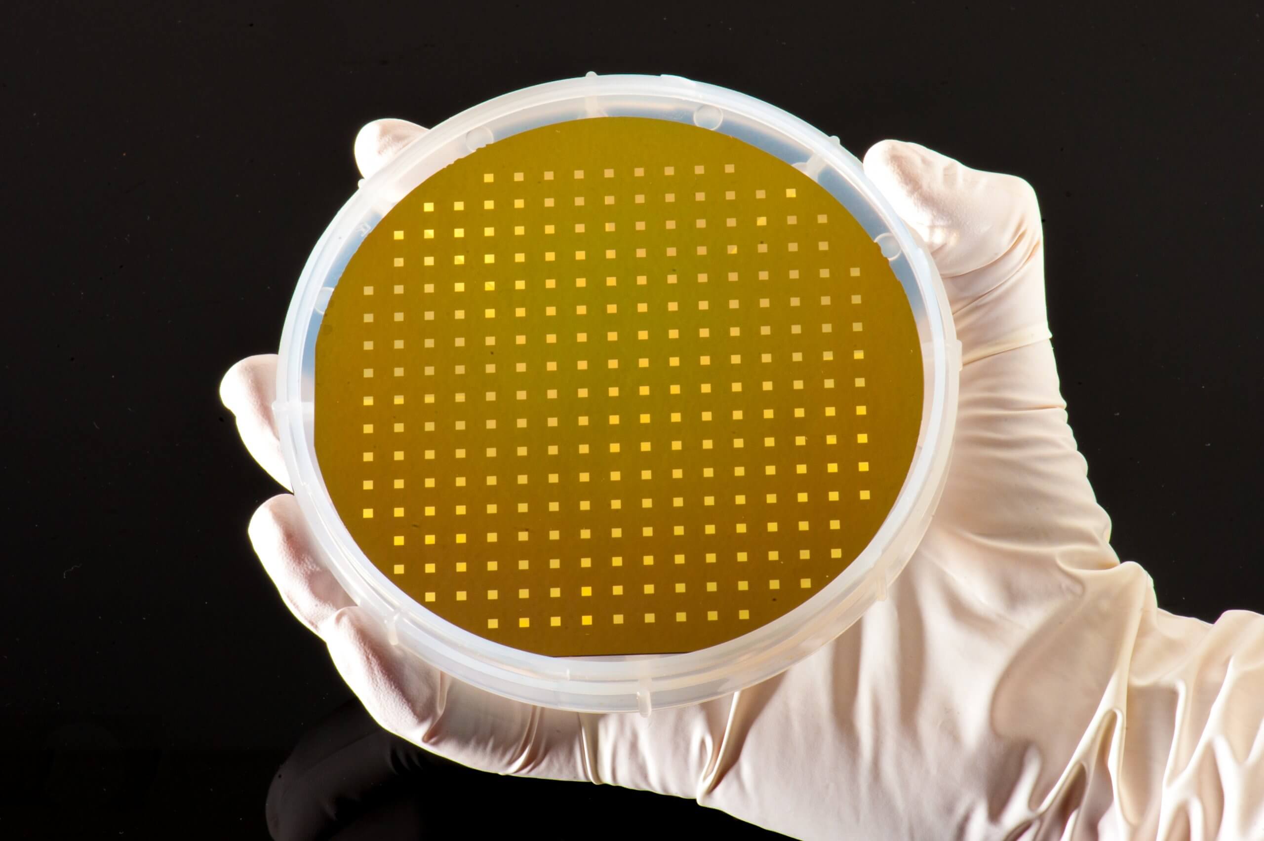
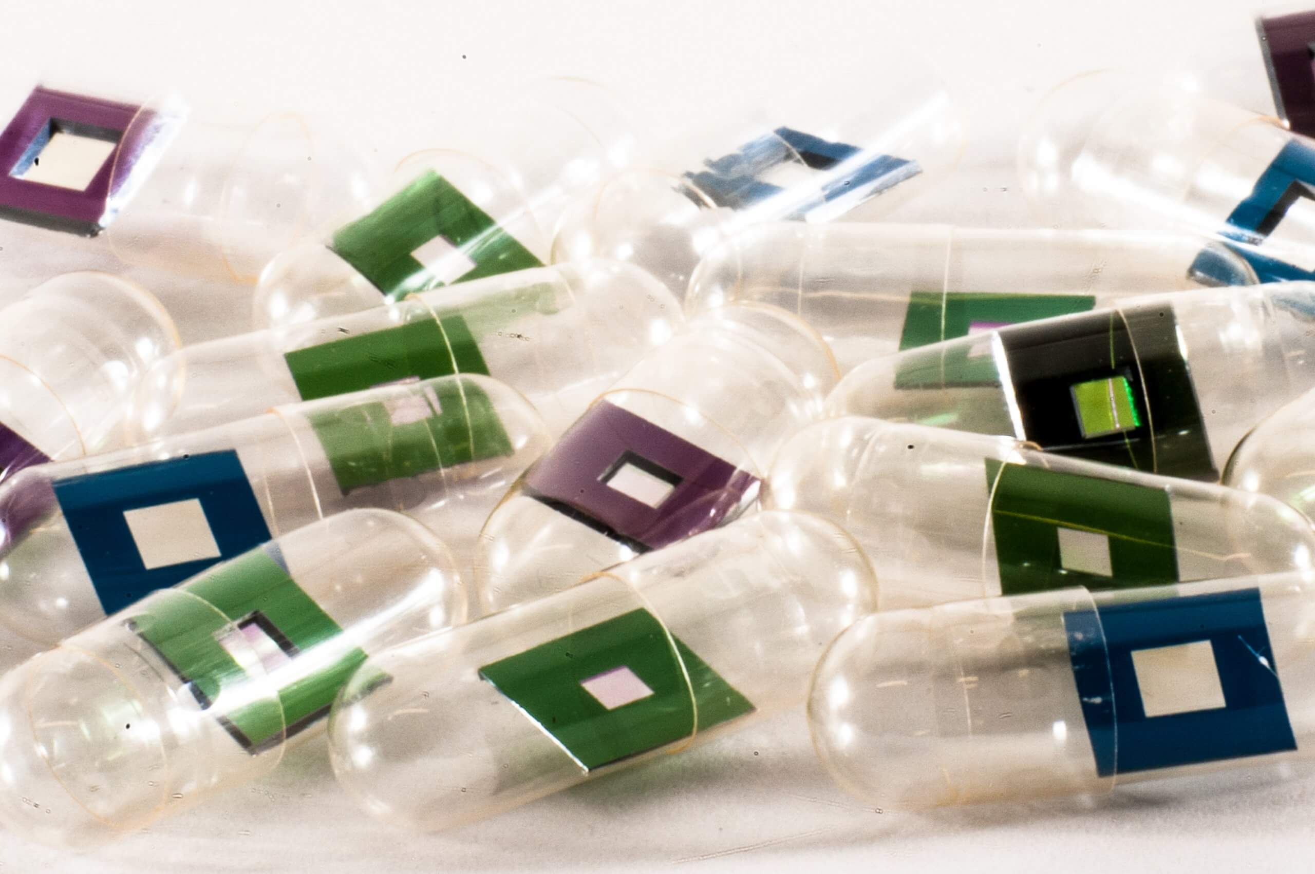
Silson’s silicon-rich nitride membranes (membrane size and thickness: 1.5 × 1.5 mm^2 and 1 µm; frame size and thickness: 5.0 × 5.0 mm^2 and 200 µm) were used for the sample preparation process and experiments were performed using the Göttingen Instrument for Nano-Imaging with X-rays (GINIX) endstation at the coherence applications beamline P10 at the PETRA III storage ring (DESY, Hamburg, Germany).
Click here to read more about this fascinating research!